2019: Easy but high-resolution live sectional imaging method of adherent cells!
“A simple microfluidic device for live-imaging of the vertical section of epithelial cells,”
Published in S. Araki, M. Nakano, M. Tsugane, F. Sunaga, M. Hattori, M. Nakano, T. Nagai, H. Suzuki Analyst, 145(2), 667-674, 2019.
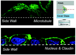
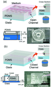
We investigated the capability of simple microfluidic devices with trenches having vertical sidewalls for live-cell fluorescence imaging of adherent cells. An epithelial cell line that forms a two-dimensional (2D) sheet was cultured to adhere to the vertical sidewall so that its vertical section can be imaged directly using ordinal inverted-type laser-scanning microscopy. The material and the structure of the device were characterized. We show that the detailed distribution of intracellular organelles, such as microtubules and mitochondria, and of intercellular apparatus, such as claudin and zonula occludens, can be imaged with high spatio-temporal resolution with a single scan.
This work has been done in collaboration with Prof. Nagai’s group.