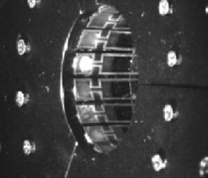
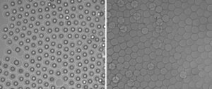
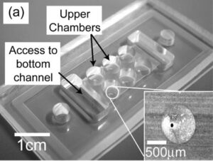
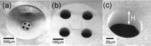
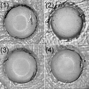
Among various single-cell analysis platforms, hydrodynamic cell trapping systems remain relevant because of their versatility. Among those, deterministic hydrodynamic cell-trapping systems have received significant interest; however, their applications are limited because trapped cells are kept within the closed microchannel, thus prohibiting access to external cell-picking devices. In this study, we develop a hydrodynamic cell-trapping system in an open microfluidics architecture to allow external access to trapped cells. A technique to render only the inside of a polydimethylsiloxane (PDMS) microchannel hydrophilic is developed, which allows the precise confinement of spontaneous capillary flow in the open-type microchannel with a width on the order of several tens of micrometers. Efficient trapping of single beads and single cells is achieved, in which trapped cells can be retrieved via automated robotic pipetting. The present system can facilitate the development of new single-cell analytical systems by bridging between microfluidic devices and macro-scale apparatus used in conventional biology.
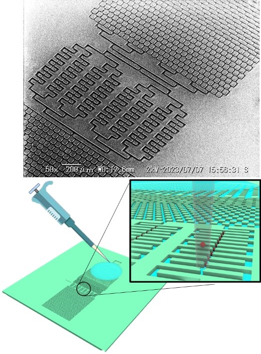
Murakami et al., Single-cell trapping and retrieval in open microfluidics, iScience, 26(11), 108323, 2023. https://www.sciencedirect.com/science/article/pii/S2589004223024008?via%3Dihub
本事業はJKA(競輪)の補助を受けて実施しました

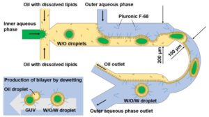
R. Ushiyama, K. Koiwai, H. Suzuki, “Plug-and-Play Microfluidic Production of Monodisperse Giant Unilamellar Vesicles Using Droplet Transfer across Water-Oil Interface,” Sens. Act. B: Chemical, 355, 131281, 2021.
Giant unilamellar vesicles (GUVs) are cell-sized compartments widely used in biological and chemical applications. The monodispersity of GUVs is essential for quantitative analysis and experimental reproducibility in biomimetic microreactor studies. In this study, we established a highly reproducible plug-and-play microfluidics-based method to generate monodisperse GUVs without surface treatment of the channel or precise flow-rate adjustment. In the microfluidic channel, we generated water-in-oil (W/O) droplets through hydrodynamic flow focusing and transferred across the W–O interface to form thin-shelled water-in-oil-in-water (W/O/W) droplets as precursors to GUVs. The success rate of the W/O droplet transfer reached nearly 100% by stabilizing the co-flow of oil and water and adjusting the curvature of the W–O interface. Owing to interfacial energy minimization, the thin oil layer of the W/O/W droplets dewetted and detached to form GUVs. We performed a membrane-protein insertion assay to confirm the unilamellarity of the GUVs and their shrinkage caused by osmotic action.
Let us know if you want to try this microfluidic device for GUV-formation.
We will send you the CAD design.
“Applying deterministic lateral displacement cell separation on immune cells of Marine shrimp”
Published in Tomoki Murakami, Keiichiro Koiwai, Hiroaki Suzuki, Sens. Act. B: Chemical, 347, 130587, 2021.
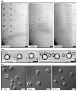
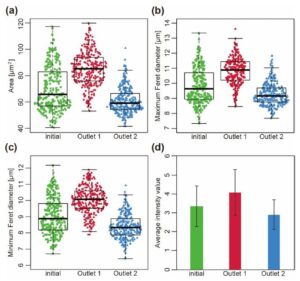
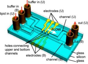
For analyzing the immune system of an organism, it is necessary to classify and separate distinct types of cells based on their characteristics. Hemocytes, immune cells of shrimp, are still difficult to classify and separate with existing technologies due to the lack of obvious morphological characteristics or type-specific markers. We adapted the deterministic lateral displacement (DLD) method as a tool to separate the hemocytes of shrimp because the previous work suggested there is a relevance between the size of hemocytes and their functions. The
image and flow cytometry (FCM) analyses confirmed that this microfluidics-based technology enabled the size-based separation of cells having a broad size distribution at the buffer condition suitable for the marine species. Cells separated with this chip retained their morphological information, and qRT-PCR was successfully conducted to reveal the differences in expression levels of immune-related genes between cell populations of different sizes. The present results prove that the DLD method can be an effective tool for the analysis of non-model organisms, in which the characteristics of cells are not well established and studied.
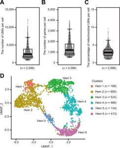
“Single-cell RNA-seq analysis reveals penaeid shrimp hemocyte subpopulations and cell differentiation process”
K. Koiwai, T. Koyama, S. Tsuda, A. Toyoda, K. Kikuchi, H. Suzuki, R. Kawano, eLife, 10:e66954, 2021.
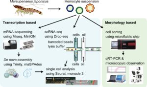
Crustacean aquaculture is expected to be a major source of fishery commodities in the near future. Hemocytes are key players of the immune system in shrimps; however, their classification, maturation, and differentiation are still under debate. To date, only discrete and inconsistent information on the classification of shrimp hemocytes has been reported, showing that the morphological characteristics are not sufficient to resolve their actual roles. Our present study using single-cell RNA sequencing revealed six types of hemocytes of Marsupenaeus japonicus based on their transcriptional profiles. We identified markers of each subpopulation and predicted the differentiation pathways involved in their maturation. We also predicted cell growth factors that might play crucial roles in hemocyte differentiation. Different immune roles among these subpopulations were suggested from the analysis of differentially expressed immune-related genes. These results provide a unified classification of shrimp hemocytes, which improves the understanding of its immune system.
“Elucidating the Membrane Dynamics and Encapsulation Mechanism of Large DNA Molecules Under Molecular Crowding Conditions Using Giant Unilamellar Vesicles“
Published in M. Tusgane & H. Suzuki, ACS Synth. Biol. 2020, 9, 10, 2819–2827.
The conservation throughout evolution of membrane-bound structures that encapsulate genomic material indicates the existence of a simple, physical mechanism that facilitates the enclosing of long-stranded DNA by lipid bilayers. This study aimed to elucidate such a mechanism by investigating how molecular crowding promotes the spontaneous enveloping of model DNA into lipid bilayer membranes. Using fluorescence microscopy and giant unilamellar vesicles (GUVs) we showed that a 166 kb DNA molecule coencapsulated with a model crowder attaches to the inner membrane of the GUVs as they osmotically deflate and after the DNA–membrane complex buds out. The set of results is consistent with the hypothesis that the depletion volume effect is responsible for the spontaneous encapsulation of DNA in the GUVs. This phenomenon may offer novel insights into the basic mechanisms governing membrane encapsulation of long-stranded nucleic acids found in celluar sytems that are independent of genetic control.
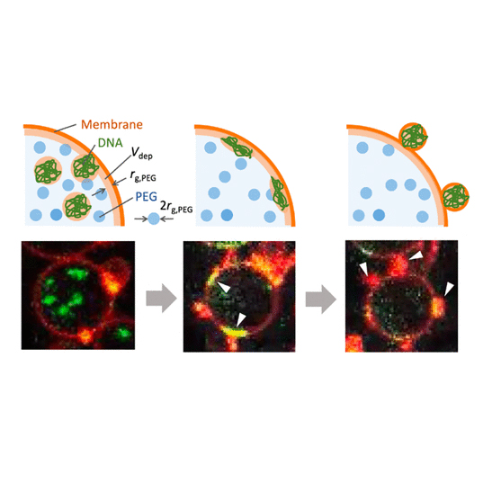
Genome-size DNA (green) spontaneously wrapped by a lipid membrane (red)
Spontaneously dividing vesicle containing genome-size DNA
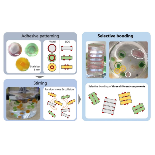
“Selective self-assembly of three-component system based on hydrophilic/hydrophobic patterning”
Published in T. Okuyama et al., Sensors and Actuators A: Physical, 312, 11214, 2020.
This paper presents a demonstration of the selective bonding of three different millimeter-scale components by using complementary patterning of hydrophilic and hydrophobic surfaces. The hydrophobic surface of millimeter-scale components fabricated from hydrophobic polydimethylsiloxane (PDMS) was made partially hydrophilic with a designed pattern by exposure to an excimer light through a stencil mask. The oil, as an adhesive, was only deposited on the hydrophobic surface. We prepared 30 components with 3 patterns, which were agitated in water by using a computer-controlled propeller stirrer. As a result, we obtained a significantly higher yield of correct bonds compared to erroneous bonds under an appropriate stirring condition. The result can be explained through the contrast in bonding strength between the correct and erroneous bonds. The design principle of using concentric patterns serves as a basic strategy for realizing programmed self-assembly with selective bonding patterns.
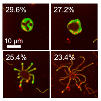
“Deformation Dynamics of Giant Unilamellar Vesicles in the Large Surface-to-Volume Ratio Regime: The Emergence of Neuron-like Morphology”
Published in K. Koseki & H. Suzuki, Langmuir 2020, 36, 22, 6238–6244.
Deformation of liposomes, or lipid vesicles, has been investigated extensively in terms of the thermodynamic equilibrium of the bending energy of the lipid bilayer membrane. However, the range of such deformation in previous literature has been limited within the moderate surface-to-volume ratio of the vesicles, in which axisymmetric shapes are dominant. Here, we show that neuron-like morphology, in which many lipid tubes extend radially from the mother vesicle, becomes dominant upon the slow osmotic shrinkage of giant unilamellar vesicles (GUVs) initially larger than several tens of micrometers. We show that, in the time-lapse confocal imaging, the emergence of lipid tubes is initiated from the instability that appeared along the annular rim of the flat stomatocyte shape. Since these deformation dynamics into the neuron-like morphology resemble that of the milk-crown formation in liquid splashing, we discuss that the Rayleigh–Plateau capillary instability drives this transformation into a nonaxisymmetric shape.
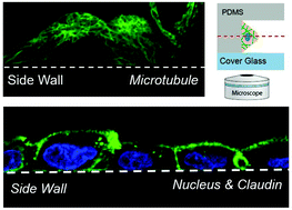
“A simple microfluidic device for live-imaging of the vertical section of epithelial cells,”
Published in S. Araki, M. Nakano, M. Tsugane, F. Sunaga, M. Hattori, M. Nakano, T. Nagai, H. Suzuki Analyst, 145(2), 667-674, 2019.
We investigated the capability of simple microfluidic devices with trenches having vertical sidewalls for live-cell fluorescence imaging of adherent cells. An epithelial cell line that forms a two-dimensional (2D) sheet was cultured to adhere to the vertical sidewall so that its vertical section can be imaged directly using ordinal inverted-type laser-scanning microscopy. The material and the structure of the device were characterized. We show that the detailed distribution of intracellular organelles, such as microtubules and mitochondria, and of intercellular apparatus, such as claudin and zonula occludens, can be imaged with high spatio-temporal resolution with a single scan.
This work has been done in collaboration with Prof. Nagai’s group.
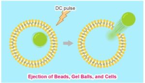
“Ejection of large particulate materials from giant unilamellar vesicles induced by electropulsation”
Published in S. Katsuta, T. Okano, K. Koiwai, H. Suzuki, Langmuir, 35(40), 13196-13204, 2019.
Electroporation or electropermealization is a technique to open pores in the lipid bilayer membrane of cells and vesicles transiently to increase its permeability to otherwise impermeable molecules. However, the upper size limit of the materials permeable through this operation has not been studied in the past. Here, we investigate the size of the material that can be released (ejected) from giant unilamellar vesicles (GUVs) upon electrical pulsation. We confirm that the volume of GUV shrinks in a stepwise manner upon periodical pulsation, in accordance with previous studies. When the same operation is applied to GUVs that encapsulate microbeads, we find that beads as large as 20 μm can be ejected across the membrane without rupturing the whole GUV structure. We also demonstrate that functional bioactive particulate materials, such as gel balls, vesicles, and cells can be encapsulated in and ejected from GUVs. We foresee that this phenomenon can be applied to precisely regulate the time and location of release of these particulate materials in the microenvironment.
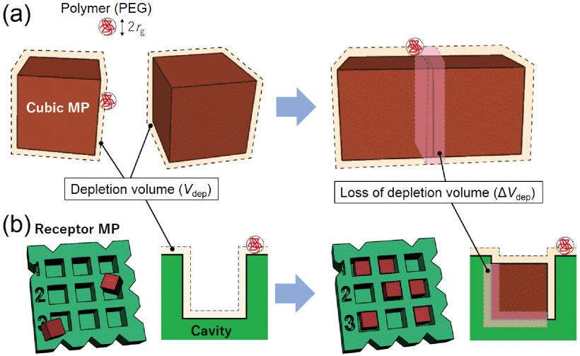
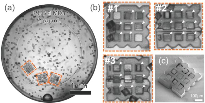
“Polymer-Induced Self-Assembly of a Three-Dimensional Microfabricated Structure”
Published in R. Kawai, Y. Mori, H. Suzuki, J. MicroElectroMechanical Syst., 28(4), 678-684, 2019.
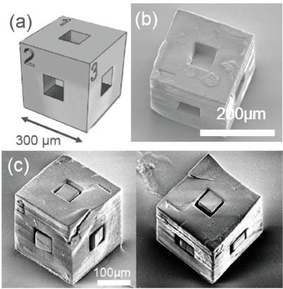
Three-dimensional (3D) self-assembly of mesoscale parts with tens to hundreds of micrometer scales, independent of the material properties, is demonstrated by simply adding an inert polymer, i.e., polyethylene glycol, with large molecular weight (4MDa) at a concentration of micrograms per milliliters. It is most likely that the depletion volume effect, a thermodynamical effect to maximize the entropy of the system, induces face-to-face bonds with microfabricated parts having flat faces. The optimum polymer concentration range yielding high assembling efficiency is found, where the bonding energy is large enough to stabilize the bonding and viscosity is small enough to allow frequent collisions. Using this strategy, a site-specific assembly of 60 μm cubic parts in cavities engraved on large receptor parts, distributed on either 2D or 3D faces, is successfully demonstrated.
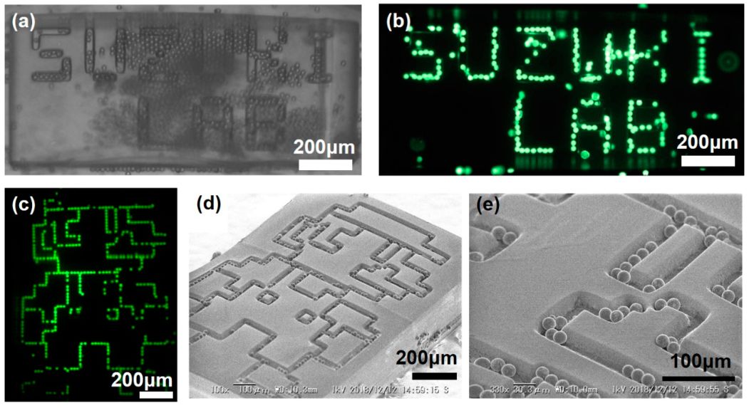
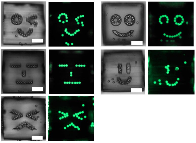
“Assembly of Microparticles to patterned trenches using the depletion volume effect”
Published in Y. Mori, R. Kawai, H. Suzuki Micromachines, 10(7), 428, 2019.
In this paper, we demonstrate that 20 μm microbeads can be preferentially assembled into substrate trenches of similar width by employing a polymer (depletant) that induces the depletion volume effect (depletion attraction). In previous works, we proved that this strategy is useful to assemble mesoscale parts in a site-specific manner. Here, we show that it is also applicable to assemble functional parts, such as fluorescent particles, into trenches engraved on the surface of two- and three-dimensional template components. A convenient advantage of this strategy is that it is independent of material properties for assembling mesoscale functional components into desired patterns.
I called up Nintendo and asked if we can use Super-Mario for scientific publication…
Lovely faces!
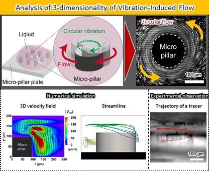
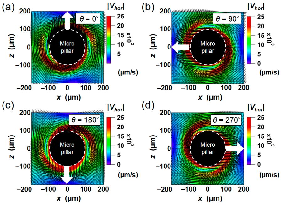
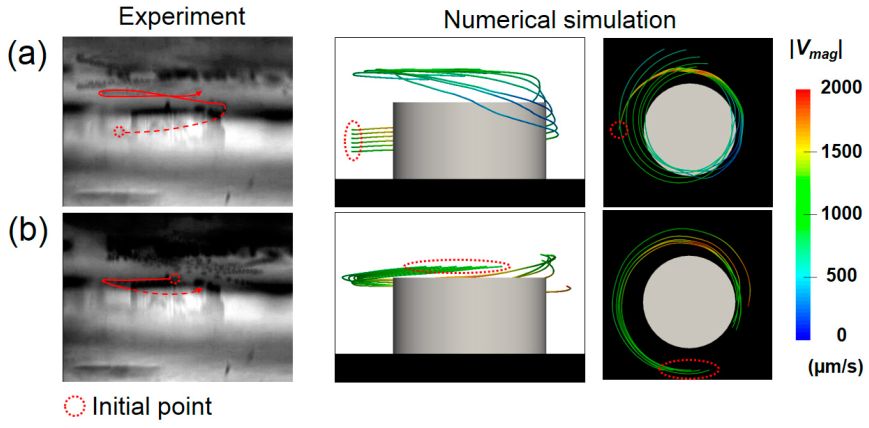
“Numerical and Experimental Analyses of Three- Dimensional Unsteady Flow around a Micro-Pillar Subjected to Rotational Vibration”
Published in K. Kaneko, T. Osawa, Y. Kametani, T. Hayakawa, Y. Hasegawa and H. Suzuki, Micromachines, 9(12), 668, 2018.
The steady streaming (SS) phenomenon is gaining increased attention in the microfluidics community, because it can generate net mass flow from zero-mean vibration. We developed numerical simulation and experimental measurement tools to analyze this vibration-induced flow, which has been challenging due to its unsteady nature. The validity of these analysis methods is confirmed by comparing the three-dimensional (3D) flow field and the resulting particle trajectories induced around a cylindrical micro-pillar under circular vibration. In the numerical modeling, we directly solved the flow in the Lagrangian frame so that the substrate with a micro-pillar becomes stationary, and the results were converted to a stationary Eulerian frame to compare with the experimental results. The present approach enables us to avoid the introduction of a moving boundary or infinitesimal perturbation approximation. The flow field obtained by the micron-resolution particle image velocimetry (micro-PIV) measurement supported the three-dimensionality observed in the numerical results, which could be important for controlling the mass transport and manipulating particulate objects in microfluidic systems.
This work was done in collaboration with Prof. Hasegawa in IIS, the University of Tokyo and Prof. Hayakawa in Chuo University!
“Selective Bonding Method for Self-Assembly of Heterogeneous Components Using Patterned Surfaces”
Published in K. Kimura, T. Okuyama, T. Okano, H. Suzuki, Sens. Act. A: Physical, 279, 306-312, 2018.
This study demonstrates the selective bonding of millimeter-scale components using the complementary patterning of hydrophilic and hydrophobic surfaces. The hydrophobic surface of the millimeter-scale components fabricated from hydrophobic polydimethylsiloxane (PDMS) was partially made hydrophilic with a designed pattern by exposure to an excimer light of 172 nm through a stencil mask. We used liquid paraffin as the adhesive and deposited it only on the hydrophobic surface. We prepared twenty components with two different complementary patterns, which were agitated in water by the computer-controlled propeller stirrer. As a result, we succeeded in obtaining a significantly higher yield of correct bonds, in comparison to that of erroneous bonds under an appropriate stirring condition. The result can be explained by the contrast in bonding strength between the correct and erroneous bonds. This principle could be used as a versatile strategy to realize programmed self-assembly with selective bonding patterns.
“Reverse Transcription Polymerase Chain Reaction in Giant Unilamellar Vesicles”
Published in M. Tsugane and H. Suzuki, Scientific Reports, 8, 9214, 2018.
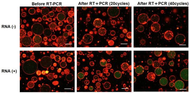
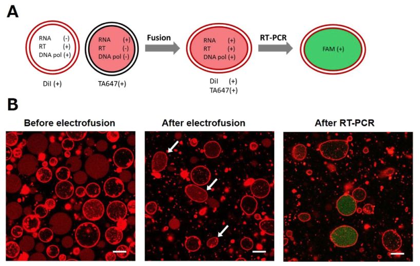
We assessed the applicability of giant unilamellar vesicles (GUVs) for RNA detection using in vesicle reverse transcription polymerase chain reaction (RT-PCR). We prepared GUVs that encapsulated one-pot RT-PCR reaction mixture including template RNA, primers, and Taqman probe, using water-in-oil emulsion transfer method. After thermal cycling, we analysed the GUVs that exhibited intense fluorescence signals, which represented the cDNA amplification. The detailed analysis of flow cytometry data demonstrated that rRNA and mRNA in the total RNA can be amplified from 10–100 copies in the GUVs with 5–10 μm diameter, although the fraction of reactable GUV was approximately 60% at most. Moreover, we report that the target RNA, which was directly transferred into the GUV reactors via membrane fusion, can be amplified and detected using in vesicle RT-PCR. These results suggest that the GUVs can be used as biomimetic reactors capable of performing PCR and RT-PCR, which are important in analytical and diagnostic applications with additional functions.
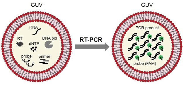
RT-PCR proceeds in GUVs so that they get green!
RT-PCR reaction can be induced by the vesicle fusion.
“A Fluidic-Based Impact Sensor”
Publishedn in D. Takahashi, K. Hara, T. Okano, H. Suzuki, PLoS One, 13(4), e0195741, 2018.
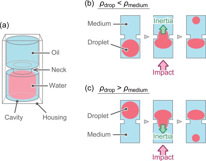
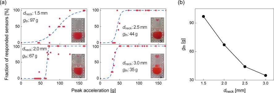
Microelectromechanical systems (MEMS)-based high-performance accelerometers are ubiquitously used in various electronic devices. However, there is an existing need to detect physical impacts using low-cost devices with no electronic circuits or a battery. We designed and fabricated an impact sensor prototype using a commercial stereolithography apparatus that only consists of a plastic housing and working fluids. The sensor device responds to the instantaneous acceleration (impact) by deformation and pinch off of a water droplet that is suspended in oil in a sensor cavity. We tested the various geometrical and physical parameters of the impact sensor to identify their relations to threshold acceleration values. We show that the state diagram that is plotted against the dimensionless Archimedes and Bond numbers adequately describes the response of the proposed sensor.
Working mechanism.
Test results.
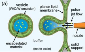
“Formation of giant vesiclelike compartments from a planar lipid membrane by a pulsed jet”
Published in K. Funakoshi, H. Suzuki, S. Takeuchi, J. Am. Chem. Soc., 129, pp. 12608-12609, 2007.
We report a straightforward method for the preparation of lipid vesicles inspired by forming soap bubbles from a soap film. Vesicles are mechanically blown out of a preformed biofunctional planar lipid membrane by applying a short pulsed liquid jet flow ejected from a fine jet nozzle brought near the membrane. This method allows us to directly encapsulate any molecules or substances of interest into the uniformly sized vesicles in a short time.
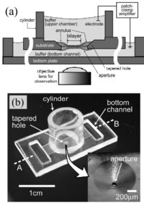
H. Suzuki, K. Tabata, H. Noji, S. Takeuchi, “PMMA micro fluidic chip for electrophysiological recording of single ion channels reconstituted in planar lipid bilayers,”Biosens. Bioelect.,22,1111-1115, 2007.
Planar lipid bilayers are used for functional studies of ion channel proteins using electrophysiological techniques. We have been developing a plastic micro-fluidic device for the reconstitution of planar lipid bilayers and electrophysiological recordings toward a “membrane protein chip” for high-throughput screening. In the previous report [Suzuki, H., Tabata, K.V., Noji, H., Takeuchi, S., 2006. Highly reproducible method of planar lipid bilayer reconstitution in polymethyl methacrylate microfluidic chip. Langmuir 22 (4), 1937–1942], we presented the method and device in which the reproducibility of planar lipid bilayers reached 90%, and multiple bilayers were formed simultaneously. In this communication, we show that our device has excellent electric properties suitable for ion channel analysis down to single molecular level. Additional aspects on the optical accessibility and controllability on lipid bilayer formation are also presented.
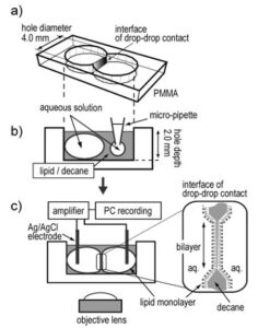
“Lipid Bilayer Formation by Contacting Monolayers in a Microfluidic Device for Membrane Protein Analysis”
Published in Kei Funakoshi, Hiroaki Suzuki, and Shoji Takeuchi, Anal. Chem. 2006, 78, 24, 8169–8174.
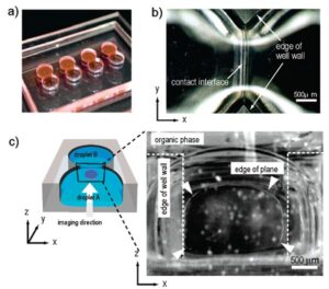
Artificial planar lipid bilayers are a powerful tool for the functional study of membrane proteins, yet they have not been widely used due to their low stability and reproducibility. This paper describes an accessible method to form a planar lipid bilayer, simply by contacting two monolayers assembled at the interface between water and organic solvent in a microfluidic chip. The membrane of an organic solvent containing phospholipids at the interface was confirmed to be a bilayer by the capacitance measurement and by measuring the ion channel signal from reconstituted antibiotic peptides. We present two different designs for bilayer formation. One equips two circular wells connected, in which the water/solvent/water interface was formed by simply injecting a water droplet into each well. Another equips the cross-shaped microfluidic channel. In the latter design, formation of the interface at the sectional area was controlled by external syringe pumps. Both methods are extremely simple and reproducible, especially in microdevices, and will lead to automation and multiple bilayer formation for the high-throughput screening of membrane transport in physiological and pharmaceutical studies.

“Highly-reproducible method for planar lipid bilayer reconstitution using a micro fluidic chip”
Published in H. Suzuki, K. Tabata, H. Noji, S. Takeuchi, “Langmuir, 22(4), pp. 1937-42, 2006.
We developed a highly reproducible method for planar lipid bilayer reconstitution using a microfluidic system made of a polymethyl methacrylate (PMMA) plastic substrate. Planar lipid bilayers are formed at apertures, 100 um in diameter, by flowing lipid solution and buffer alternately into an integrated microfluidic channel. Since the amount and distribution of the lipid solution at the aperture determines the state of the lipid bilayer, controlling them precisely is crucial. We designed the geometry of the fluidic system so that a constant amount of lipid solution is distributed at the aperture. Then, the layer of lipid solution was thinned by applying an external pressure and finally became a bilayer when a pressure of 200-400 Pa was applied. The formation process can be simultaneously monitored with optical and electrical recordings. The maximum yield for bilayer formation was 90%. Using this technique, four lipid bilayers are formed simultaneously in a single chip. Finally, a channel current through gramicidin peptide ion channels was recorded to prove the compatibility of the chip with single molecule electrophysiology.
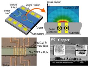
“A Chaotic Mixer for Magnetic Bead-Based Micro Cell Sorter”
Published in H. Suzuki, C. M. Ho, N. Kasagi, MicroElectroMechanical Systems, 13(5),2004, pp. 779-790.
This is the work I have mostly done in Dr. Chih-Ming Ho’s lab at UCLA. It is also my PhD work.
An efficient magnetic force driven mixer with simple configuration is designed, fabricated, and tested. It is designed to facilitate the mixing of magnetic beads and biomolecules in a microchannel, where mixing is unavoidably inefficient due to its low Reynolds number. With appropriate temporal variations of the force field, chaotic mixing is achieved, hence the mixing becomes effective. The mixing device consists of embedded microconductors as a magnetic field source and a microchannel that guides the streams of working fluid. It is demonstrated that a pair of integrated micro conductors provides a local magnetic field strong enough to attract nearby magnetic beads. Mixing of magnetic beads is accomplished by applying a time-dependent control signal to a row of conductors. Two-dimensional numerical simulation has been performed to design the configuration of the channel and
electrodes, which creates chaotic motion of beads. It is found that a simple two-dimensional serpentine channel geometry with the transverse electrodes is able to create the stretching and folding of material lines, which is a manifestation of chaos. The mixing pattern predicted by the simulation has been confirmed by both flow visualization and PTV (Particle Tracking Velocimetry) in
the chaotic mixer fabricated, which should greatly increase the attachment of beads onto the target biomolecules. The optimum frequency of applied control signal is searched by evaluating the
Lyapunov exponent in both numerical and experimental particle tracking.
“Planar lipid bilayer reconstitution with micro fluidic system”
published in H. Suzuki, K. Tabata, Y. Kato-Yamada, H. Noji, S. Takeuchi, Lab Chip, 4, 502-505, 2004.
This is my first publication for making planar lipid bilayer using a microfluidic chip. It was beginning of the Lab on a Chip journal. Good old days.
A planar lipid bilayer which is widely used for the electrophysiological study of membrane proteins in laboratories is reconstituted using a micro-fluidic system, in a manner that is suitable for automated processing. We fabricated micro-channels on both sides of the substrate, which are connected through a 100–200 μm aperture, and showed that the bilayer can be formed at the aperture by flowing the lipid solution and buffer, alternately. Parylene coating is found to be suitable for both bilayer formation and electric noise reduction. Future applications include a high-sensitivity ion sensor chip and a high-throughput drug screening device.
“Active control of an axisymmetric jet with distributed electromagnetic flap actuators”
Published in H. Suzuki, N. Kasagi, Y. Suzuki, Experiments in Fluids 36 (2004) 498–509.
This is my very first original paper.
Miniature electromagnetic flap actuators are developed and mounted on the periphery of the nozzle exit of an axisymmetric jet to induce various flow modes and enhance mixing processes. It is demonstrated that the flap actuators can significantly modify the large-scale vortical structures. In particular, when the flaps are driven in antiphase on either side of the jet, alternately inclined and bent vortex rings are generated, and the jet bifurcates into two branches.